

Introduction
Gene products exert their function in a timely and spatially defined fashion, within a specific molecular and cellular environment. Knowledge of qualitative and quantitative gene expression profiles is central to the understanding of the role and activity of gene products in complex biological processes. Monitoring global profiles of gene expression allows for defining a comprehensive molecular phenotype and to capture dynamic molecular changes associated with cell differentiation and proliferation, apoptosis, developmental stages, disease status such as cancer or response to drug and treatments. The analysis of gene expression by microarray technology is widely used in the field of cancer research and allows the parallel expression analysis of tens of thousands of genes / transcript species within a single experiment, thus providing rapid information about the transcriptional status of representative parts of the genome. Therefore, using DNA-microarray technology one can get insights into disease specific cellular regulation pathways and action / regulation of genes with unknown function. For such DNA microarray-based experiments, fluorophor-labelled cDNA pools, derived from transcripts isolated from tumor and control tissues, are hybridised to PCR amplified gene-specific cDNA fragments (cDNA-microarrays) or chemically synthesized oligonucleotides (e.g., 70mer oligonucleotide microarrays), immobilized on coated glass surfaces. The complex analysis of measured fluorescence ratios of gene-specific features and its biological / biomedical interpretation is done by sophisticated statistical and biocomputational means.
Main focus of our research was: (1) identification of unknown pathomechanisms in specific tumor entities, (2) identification of candidate genes and gene signatures suited for prognosis and stratification of patients, (3) identification of new signaling pathways with oncogenic potential.
Results/Project Status
Search for putative tumour suppressors on 1p36.31-13 and 19q13.2-33, frequently affected in oligodendroglioma
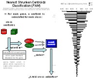
Loss of heterozygosity (LOH) on both, the short arm of chromosome 1 and the long arm of chromosome 19 on chromosomal regions 1p36.31-13 and 19q13.2-33 is highly recurrent and associated with a more favorable prognosis and better response to chemotherapy. However, the putative oligodendroglioma tumour suppressor genes on these chromosomal regions are not known yet. To further investigate those two regions in glioma, we performed gene expression profiling with cDNA microarrays comprising two continuous regions covering 1p36.31-13 and 19q13.2-33. Then, we identified the top up- and down-regulated genes in all 1p/19q cases with retention (RET) and in all 1p/19q cases with LOH, respectively. Determination of differentially expressed genes was carried out by applying Significant Analysis for Microarrays (SAM) and Nearest Shrunken Centroids method (PAM) for several combinations.
Chipdesign: cDNA microarrays consisting of 7000 gene specific cDNAs, each spotted in triplicate; in addition, region specific cDNAs: 1p36.31-13: 118 cDNAs, 19q13.2-33: 96 cDNAs; tumour RNA was co-hybridized with commercially available reference RNA (Stratagene), composed of total RNA from ten human cancer cell lines for all tumour samples; colour switch experiments were performed.
Sample collective: 10 cases of grade II diffuse astrocytoma; 7 cases of grade II low grade oligodendroglioma; 14 cases of grade III anaplastic oligodendroglioma: 7 cases of grade III anaplastic oligoastrocytoma.
Strategies: Identification of:
- top up-/ down-regulated genes in 1p/19q RET and 1p/19q LOH cases (ln x > 1; ln x < –1)
- gene expression differing in 1p/19q RET and 1p/19q LOH cases (> ln 0.6; < ln –0.6)
- significant differentially expressed genes (via SAM)
- genes best discriminating (via Nearest Shrunken Centroid analysis) between:
- LOH1p/19q (all tumours) versus no LOH1p/19q (all tumours)
- LOH1p/19q (WHO grade III) versus no LOH1p/19q (WHO grade III)
- LOH1p/19q (WHO grade II) versus no LOH1p/19q (WHO grade II)
- all oligodendroglioma (WHO grade II) versus all anaplastic oligodendroglioma (WHO grade III)
- oligodendroglioma (WHO grade II, with LOH 1p/19q) versus anaplastic oligodendroglioma (WHO grade III, with LOH1p/19q)
We identified about 10 genes which are able to significantly discriminate between the subclasses, some of them specific for different grading of glioma which might suite as tumour suppressors. We found a further gene, which was promotor-hypermethylated in case of LOH 1p/19q. Hypermethylation of this gene is a significant predictor of longer survival in
patients with oligodendroglial tumors. Quantitative real-time reverse transcription-PCR analyses were performed for several candidate genes and confirmed the microarray findings. Taken together, our study provides a set of interesting novel candidate genes that may play important roles in the pathogenesis of oligodendroglial tumors.
A minimal set of transcripts characterizing glioblastoma multiforme long-term survivors (LTS)
Glioblastoma multiforme (GBM, WHO grade IV) is the most malignant and also the most frequent astrocytic tumour, composed of poorly differentiated neoplastic astrocytes. It accounts for approximately 12-15% of all intracranial neoplasms and 50-60 % of all astrocytic tumours with a median survival < 1 year. However, a small subgroup of GBM patients has a better clinical outcome, with a small number of patients surviving several years. To investigate the molecular differences between long term surviving (LTS) and short term surviving (STS) patients with GBM we performed expression profiling on 12 LTS and 12 STS patients, followed by a classification with two independent clustering methods: shrunken centroid analysis (SCA) and significant analysis of microarrays (SAM). We identified a set of genes differentially expressed in the two subclasses of GBM. The most promising candidate genes are currently analyzed in more detail for its function and regulation.
RNA expression profiling of gliomas
We are analyzing the comprehensive RNA expression profiles of the 96 primary glioma constituting the “Glioma Core Collection” of the NGFN2 Brain Tumor Network. For this purpose, a 35,035 oligonucleotide microarray representing approximately 25,100 unique genes and 39,600 transcripts, was generated for the analyses of brain tumors. The microarray was complemented by 500 additional 70mer probes, which are specific for genes in the commonly deleted chromosomal regions 10q24-qter and 22q. Currently, we are performing a specific RNA amplification procedure [4] for all RNA samples, hybridization of samples on 35k oligonucleotide micraoarrays and primary data analysis.
Outlook
We identified a distinct set of genes that are located within previously defined candidate tumor suppressor gene regions on 1p36.22-p36.31 and 19q13.3 and showed significantly lower transcript levels in 1p/19q-deleted gliomas as compared to 1p/19q-intact gliomas. We additionally found novel oligodendroglioma progression-associated candidate genes whose expression differed significantly between WHO grade II and WHO grade III oligodendrogliomas. Further studies are needed to elucidate the molecular mechanisms underlying the differential expression of these newly identified candidate genes and to clarify their functional roles in oligodendroglial tumors. Further functional studies will also be done in case of glioblastoma multifome LTS candidate genes.
Our work on Oligodendroglioma and GBM LTS demonstrates that RNA expression profiling is a powerful technique capable of identifying clinically relevant molecular markers and putative therapy target genes.
Lit.: 1. Wrobel et al. Microarray-based gene expression profiling of benign, atypical and anaplastic meningiomas identifies novel genes associated with meningioma progression. Int J Cancer. 2005 Mar 20;114(2):249-56. 2. Neben et al. Microarray-based screening for molecular markers in medulloblastoma revealed STK15 as independent predictor for survival. Cancer Res. 2004 May 1;64(9):3103-11. 3. Korshunov et al. Gene expression patterns in ependymomas correlate with tumor location, grade, and patient age. Am J Pathol. 2003 Nov;163(5):1721-7. 4. Schlingemann J et al. Effective transcriptome amplification for expression profiling on sense-oriented oligonucleotide microarrays. Nucleic Acids Res. 2005 Feb 17;33(3):e29.