

Introduction
The role of genetic factors in the pathogenesis of Parkinson’s disease (PD) has been discounted for a long time. This reflects a variety of factors complicating the identification of genetic factors in PD like late onset of the disease, reduced penetrance of genetic traits, genetic heterogeneity and environmental factors contributing to the disease. First systematic approaches to define a genetic contribution to PD focused on twin studies, but remained inconclusive for several reasons. A major contribution to the identification of genetic factors in the pathogenesis of typical PD came from linkage studies in large families with autosomal dominant or autosomal recessive inheritance of typical Parkinsonism to date 11 chromosomal loci were identified harbouring disease-causing genes (PARK1-11, Online Mendelian Inheritance in Man; http://www.ncbi.nlm.nih.gov/). The identification of mutations in five of these genes and subsequent functional characterization of these disease genes allowed insight into molecular pathways leading to neurodegeneration and dysfunction of the nigrostriatal system. There is increasing evidence that genes involved in monogenic forms of the disease may act as susceptibility factors also in the common sporadic form of PD. Thus disturbance of the ubiquitin proteasome pathway came into focus of interest and underscored the relevance of protein misfolding and accumulation in neurodegeneration in PD. Other genes, like DJ-1 and PINK1, are involved in mitochondrial homeostasis and linked newly identified signalling pathways to the established paradigm of oxidative stress in PD. Today it is generally accepted, that apart from familial forms of PD that account for approximately 20% of the disease, environmental factors acting on genetically predisposed individuals play a role in the majority of sporadic PD patients. In the our studies we search for novel genetic susceptibility factors and investigate molecular signalling pathways of these and known disease genes in cell culture in vitro and in animal models in vivo to define their role in neurodegeneration. To achieve this goal, cellular and animal models are studied with focus on the protein degradation machinery (ubiquitination, aggregation), mitochondrial homeostasis (oxidative stress, energy depletion), characterization of existing animal models (pathological and behavioural dissection of transgenic animals over-expressing wild-type and disease-associated mutant synphilin-1) and transcriptome analysis using Affymetrix microarray analysis.
Results/Project Status
Omi/HtrA2 in Parkinson’s disease
The serine protease Omi/HtrA2 is a nuclear encoded protein with a mitochondrial targeting sequence. Omi/HtrA2 is released from the mitochondria into the cytosol during apoptosis and initiates caspase-dependent cell death via inhibition of inhibitor of apoptosis proteins and caspase-independent cell death via its serine-protease activity. Moreover Omi/HtrA2 belongs to a family of PDZ-domain serine proteases that exhibit a chaperone-like function. A first link to neurodegeneration was established by its identification as a presinilin-1- and amyloid-beta-interacting protein - both proteins are involved in the pathogenesis of inherited Alzheimer’s disease. Recently Omi/HtrA2-knockout mice have been generated that show a neurodegenerative phenotype with Parkinsonian symptoms suggesting a neuroprotective role of the serine protease activity of Omi/HtrA2 in vivo. Using a candidate gene approach on genes involved in cellular stress response and protein misfolding and mitochondrial homeostasis we identified two novel mutations in the Omi/HtrA2 gene in sporadic PD patients [1]. In four German PD patients a heterozygous G399S substitution in the PDZ domain was identified, that was absent in a total of 740 controls. Moreover a novel polymorphic amino acid variant of Omi/HtrA2 that was associated with PD was found. Both mutations markedly reduced the activation of protease activity of Omi/HtrA2 when compared to the wild-type protein. Immunohistochemistry and functional analyses in stably transfected cells revealed that G399S mutant Omi and to a lesser extend the risk allele of the newly defined A141S polymorphism induced mitochondrial swelling and mitochondrial membrane dysfunction. Viability assays revealed that cells overexpressing PDZ mutant Omi/HtrA2 were more susceptible to staurosporine-induced cell death than wild type [1]. Based on functional genomics these results on mutations in the Omi/HtrA2 gene in PD provide a novel link between mitochondrial dysfunction and the neurodegeneration in PD. Therefore these results and data on PD-related genes PINK1 and DJ-1 suggest an important role of nuclear encoded proteins that are targeted to mitochondria conferring protection in terms of mitochondrial stress.
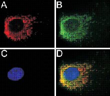
Synphilin-1 and its interacting proteins
We recently identified a missense mutation in two sporadic Parkinson patients in synphilin-1, a protein of unknown function which interacts with alpha-synuclein. In order to further elucidate the pathogenesis of nigral degeneration, we searched for novel synphilin-1 interacting proteins by yeast two-hybrid screening and deciphered the highly insoluble protein periphilin as new interactor. Subsequent immunohistochemical studies in brains of PD patients identified periphilin as a component of Lewy bodies. As periphilin maps to the PARK8 region, we searched for mutations in familial PD. In two affected patients of a small family compatible with autosomal dominant inheritance, a 205A>G sequence variant leading to a K69E exchange was detected. To further characterize wild type and mutant periphilin, we performed immunocytochemical and functional analyses. We show that periphilin localizes to the nucleus. The K69E exchange does neither affect the cellular distribution nor the susceptibility to cellular stressors such as the proteasome inhibitor MG132 or H2O2. However, human embryonic kidney cells stably expressing mutant periphilin appeared to be more susceptible to the nitric oxide donor S-nitroso-N-acetylpenicillamin (Glass et al. submitted). Since periphilin accumulated under proteasomal inhibition with MG132 it seems likely that this protein is degraded by the proteasome. However, neither ubiquitinitation nor sumoylation as protein degradation signals could be proven. Together with the group of Prof. Wurst a knock out model of periphilin was generated. These mice, however, are not viable but serve as a valuable tool to fine map the expression of periphilin as the LacZ gene has been integrated into the periphilin gene as a marker. These mice clearly show that periphilin is indeed expressed in the substantia nigra, a brain region which is most prominently affected in Parkinson’s disease. In summary, our results link periphilin and synphilin-1 and open up new insights for periphilin as a novel potential pathogenetic factor in PD.
Outlook
Substantial progress in the understanding of the molecular signalling pathways in the pathogenesis of PD came from the identification of genetic factors in inherited forms of the disease. Based on functional analyses of the encoded proteins the ubiquitin-mediated protein degradation machinery came into focus of interest. Genes mutated in inherited forms of Parkinsonism encode proteins that are able to interfere with proteasomal function at different stages: (i) due to degradation via the ubiquitin-proteasome system (alpha-synuclein, Parkin, mutant DJ-1) or as integral components of the degradation pathway (Parkin, UCH-L1). Subsequently variants acting as susceptibility factors for sporadic PD could be identified in all of these genes. The functional characterization of PINK1 and DJ-1 revealed their physiological role in oxidative stress response and mitochondrial function and offered the possibility to link previously identified biochemical alterations in sporadic PD, i.e. complex I deficiency and mitochondrial dysfunction, with genetic risk factors. In our studies we provided further evidence for a relationship between mitochondrial dysfunction and protein misfolding in the pathogenesis of PD. Thus the identification and characterization of susceptibility genes supports the idea of a heterogeneous aetiology of PD and indicate first steps towards a unifying concept in the pathogenesis of PD.
To further elucidate the pathogenesis of PD we are currently characterizing novel interactors of synphilin-1. Also, constructs to generate knock out mice of synphilin-1, a project which is performed together with the group of Prof. Wurst, are ready for generating ES cells. It will be interesting to learn if these mice are (i) viable, (ii) have signs of neurodegeneration, and/or (iii) have an altered dopamine pathway.
Lit.: 1. Strauss KM et al. Loss of function mutations in the Omi/HtrA2 gene in Parkinson’s disease. Hum Mol Genet. 2005 Aug 2; 14, 2099-2111. 2. Glass A et al. Periphilin is a new interactor of synphilin-1, a protein involved in Parkinson’s disease. Submitted.