


Introduction
Radiation and chemotherapy, as well as the surgical removal of tumour tissue, are the classical weapons in the fight against cancer. Although these methods are still in use, they often do not lead to sustainable success. Therefore, combatting cancer by activating the immune system seems to be an auspicious strategy for the development of new cancer treatments. Absolutely essential for successful cancer treatment is the identification of tumour associated antigens (TAA) against which the immune system can be targeted. Proteins expressed at low levels in normal tissue, but high in the tumour, build one class of tumour associated antigens. The vital parameter for immunotherapies based on the effects of cytotoxic T cells (CTLs) is the epitope concentration as far as overexpression is concerned. Epitope concentration means the relative frequency of one specific MHC-peptide complex on the tumour surface. Extensive studies for such frequency ratios of MHC ligands on tumours have not been undertaken so far. Our laboratory recently developed a method enabling us to measure exact quantitative differences of MHC-bound peptides between normal and tumour tissue.
The aim of this project combining genomics and functional proteomics is to show a possible connection between the MHC-peptide complex density and the mRNA expression of the source proteins of those peptides, especially between tumour and normal tissue. No significant correlation would challenge seriously all approaches using overexpressed tumour antigens based on gene expression data and therefore make our method for direct quantification of MHC ligands indispensable. If there is a significant correlation between MHC peptide density and gene expression, our project would essentially contribute clarifying the quantitative context between a cell�s transcriptome and the entirety of presented HLA ligands, known as ligandome.
Beyond that, the findings would also give a solid experimental basis for applying the very efficient applicable DNA microarray technology for the identification of tumour-associated CTL epitopes. Results during research and final results can be transferred directly to ongoing clinical studies for experimental cancer immunotherapy.
Project Status
State-of-the-art
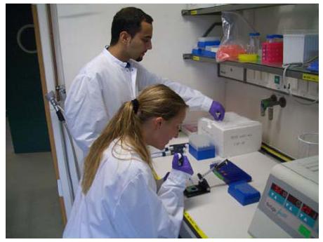
Fig 1: Junior scientists handling RNA probes for gene expression analysis
Proteins overexpressed in cancer patients provide a source for potential tumour-associated CTL epitopes. Prominent examples include HER-2/neu1,2, MUC13, CEA2 or p534. Despite their lack of absolute tumour-specificity, MHC-bound peptides from those proteins were shown to elicit T cell responses directed against tumours in vitro as well as in clinical trials. Therefore, there is sufficient evidence to see the search for overexpressed antigens as a potentially successful method of cancer immunotherapy. Overexpression of TAA mostly refers to the protein or mRNA level, usually measured with antibodies, PCR or microarrays. However, since CTLs recognize peptides bound to MHC molecules on the target cell surface, the density of such complexes bearing the peptide of interest will be the key determinant for defining overexpression in this system. Indeed, epitope density was reported to be an important parameter for T cell priming5,6 as well as for target cell lysis in the CTL effector phase7.
Project Status
For more than ten years we have been isolating and analyzing MHC-bound peptides from cell lines and tissue samples. This led to the characterization of more than 50 allele-specific peptide motifs. As an application of such peptide motifs, we established prediction algorithms for T cell epitopes accessible to the public by our internet database �SYFPEITHI�8. For the identification of peptides, we now rely mostly on sequencing by tandem mass spectrometry (MS). We have developed a technique to detect and verify predicted potential T cell epitopes9 which resulted in the identification of a new epitope from the tumour antigen MAGE-A110. We have started to pursue a strategy to identify as many candidates as possible for tumour-associated T cell epitopes in individual patients11. In this approach, we combine MHC-ligand identification with gene expression analysis of individual tumours to identify peptides corresponding to overexpressed or exclusively expressed genes in the tumour. For this project, we established the Affymetrix GeneChip® technology in our laboratory four years ago and have gained sample experience with it12. Potential epitopes identified by this combined approach are being directly transferred into clinical vaccination studies against renal cell carcinoma. Some peptides have already been verified as true T cell epitopes mediating tumour cell lysis13. We are currently able to routinely identify 100 MHC ligands from one tumour sample and have recently succeeded in developing a method that for the first time allows the direct and quantitative comparison of MHC presented peptides from sample pairs, such as tumour and corresponding healthy tissue by online liquid chromatography mass spectrometry (LCMS) 14.
During the time of commencement, establishment and optimization of essential techniques was to the fore. After extensive experimentation a protocol has been designed for reproducible differential quantitative analysis of HLA-ligands from human primary samples. Gene expression analysis with oligonucleotide-microarrays essential for these analyses has also been established. The first human renal cell carcinomas has been processed and the number of identified peptides are in accordance with our expectations.

Fig 2: Professor Dr. Hans-Georg Rammensee
The essential scientific conclusion of this project will be to verify whether there is a direct connection between transcriptome and HLA ligandome or not. The data will admit a substantiated appraisal, independently of the result, how reasonable the definition of tumour associated cell antigens using overexpression on mRNA level is. Research regarding the antigen processing and antigen presentation, the two steps between transcriptome and ligandome, can also benefit from these quantitative connections. As a direct scientific application, tumour antigens identified in this project are used in a clinical cancer immunotherapy trial in renal cell carcinoma patients. Antigen processing, i.e. the steps leading from proteins to MHC ligands, is still an area of intensive research, also in our group. One important question refers to whether MHC ligands are generated by the normal protein turnover of cells or mainly from defective ribosomal products. A quantitative correlation of MHC ligands with their encoding mRNA might provide new insights into this question as well as into other steps of antigen processing. Independent of such intermediate steps, knowing the correlation between transcriptome and MHC ligandome will be extremely important in the search for overexpressed tumour-associated antigens. A close correlation would support the current approach employed by many groups to define overexpression of tumour antigens based on mRNA expression, even though a solid scientific basis for this has as yet been lacking. If, on the other hand, no such correlation is observed, our method for the differential MHC ligand quantification will become the only feasible tool for screening large numbers of new, overexpressed tumour antigens. Independent of the outcome, we expect the MHC ligand analysis to reveal new antigens unique to tumors that cannot be detected by microarrays, such as for example peptides derived from mutated genes, altered mRNA splicing or even protein splicing.
Lit.: 1. Fisk B. et al. Identification of an immunodominant peptide of HER-2/neu protooncogene recognized by ovarian tumor-specific cytotoxic T lymphocyte lines. J. Exp. Med. 181, 2109-2117 (1995). 2. Kawashima I. et al. Identification of HLA-A3-restricted cytotoxic T lymphocyte epitopes from carcinoembryonic antigen and HER-2/neu by primary in vitro immunization with peptide-pulsed dendritic cells. Cancer Res. 59, 431-435 (1999). 3. Brossart P. et al. Identification of HLA-A2-restricted T-cell epitopes derived from the MUC1 tumor antigen for broadly applicable vaccine therapies. Blood 93, 4309-4317 (1999). 4. Vierboom M.P. et al. Tumor eradication by wild-type p53-specific cytotoxic T lymphocytes. J. Exp. Med. 186, 695-704 (1997). 5. Wherry E.J. et al. The induction of virus-specific CTL as a function of increasing epitope expression: responses rise steadily until excessively high levels of epitope are attained. J. Immunol. 163, 3735-3745 (1999). 6. Bullock T.N. et al. Antigen density presented by dendritic cells in vivo differentially affects the number and avidity of primary, memory and recall CD8+ T cells. J. Immunol. 170, 1822-1829 (2003). 7. Rawson P. et al. Immunotherapy with dendritic cells and tumor major histocompatibility complex class I-derived peptides requires a high density of antigen on tumor cells. Cancer Res. 60, 4493-4498 (2000). 8. Rammensee H.G. et al. SYFPEITHI: database for MHC ligands and peptide motifs. Immunogenetics 50, 213-219 (1999). 9. Schirle M. et al. Identification of tumor-associated MHC class I ligands by a novel T cell-independent approach. Eur. J. Immunol. 30, 2216-2225 (2000). 10. Pascolo S. et al, A MAGE-A1 HLA-A A*0201 epitope identified by mass spectrometry. Cancer Res. 61, 4072-4077 (2001). 11. Weinschenk T. et al. Integrated functional genomics approach for the design of patient-individual antitumor vaccines. Cancer Res. 62, 5818-5827 (2002). 12. Schoor O. et al. Moderate degredation does not preclude mircoarray analysis of small amounts of RNA. Biotechniques 35, 1192-1201 (2003). 13. Schmidt S. et al. Induction of adipophilin-specific cytotoxic T lymphocytes using a novel HLA-A2-binding peptide that mediates tumor cell lysis. Cancer Res. 64, 1164-1170 (2004). 14. Lemmel C. et al. Differential quantitative analysis of MHC ligands using stable isotope labelling in mass spectrometry. Nat. Biotechnol. 22, 450-454 (2004).