Introduction
Stroke is a devastating neurological disease. It is the third most common cause of death and enduring disability in the western world. Treatment with thrombolysis is limited by side effects and can be applied to less than 10 percent of patients. Therefore, basic research focuses on other therapeutic approaches than restoration of blood circulation. Although stroke is characterized by a sudden onset of symptoms, it also triggers delayed processes that markedly determine the functional and morphological outcome. Delayed events are angiogenesis, neurogenesis, inflammation, and apoptosis. All these events rely heavily on gene transcription. Characterization of gene expression in stroke holds the promise to identify new mechanisms that may be targeted by small molecule drugs. Several techniques have been developed to investigate gene transcription on a large scale. All approaches have advantages and pitfalls. No single technique will produce a complete list of induced genes. Therefore, we have employed several techniques in parallel.
Results/Project Status
To profile gene expression after focal cerebral ischemia we had developed a variant of differential display PCR called restriction-mediated differential display. Using this technique we have analysed gene expression after transient and focal cerebral ischemia (1, 2). Two genes found to be upregulated by ischemia were further investigated. Both turned out to be pro-apoptotic in function. Because of the limitations inherent in any technique we also applied microarrays and a signature-based method to the analysis of ischemia-induced gene expression.

MPSS…
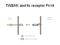
Massively parallel signature sequencing (MPSS) after focal cerebral ischemia and RNA extraction from the whole hemisphere showed that the cytokine TWEAK is upregulated after cerebral ischemia. We confirmed the induction of TWEAK by RT-real time PCR and found that in addition the membrane receptor of TWEAK, Fn14, was upregulated even more profoundly. An in vitro ischemia model, oxygen glucose deprivation, also stimulated TWEAK and Fn14 expression and provided evidence that the expression is localized in neurons. The presence of the membrane receptor Fn14 and its upregulation in neurons raised the question of what the effect of TWEAK would be on neurons. We have, therefore, exposed primary cortical neurons to recombinant TWEAK and found an increased apoptosis rate. TWEAK is known to activate the transcription factor NF-#B. Indeed, TWEAK stimulated NF-#B also in neurons. Inhibition of NF-#B ameliorated TWEAK-induced apoptosis showing that NF-#B mediates the pro-apoptotic effect of TWEAK. In an in vivo-model of cerebral ischemia a neutralizing antibody against TWEAK reduced the infarct size. All these data suggest that TWEAK, which we have found by MPSS, is a cytokine that mediates the ischemic brain damage. Our data have been published in 2004 (3).
Outlook
Previous approaches to characterize gene expression after focal cerebral ischemia failed to differentiate between neurons, glia, and endothelial cells. Especially rare cell populations such as new born neurons are likely to be underrepresented in whole brain extracts.
Focal and global cerebral ischemia stimulates proliferation of adult neural progenitor cells in the subventricular and the subgranular zone. Adult neural progenitor cells are characterized by the expression of nestin. New born neural cells leave usual migratory pathways and immigrate into the ischemic cortex or striatum, where they differentiate into astrocytes and neurons. Although the number of neurons replaced after ischemia remains small without further treatment, administration of growth factors such as EGF stimulated neuronal replacement 100-fold. Under these conditions more than 20 % of neuronal subpopulations lost after ischemia were replaced. Evidence that new born are functionally incorporated in neuronal networks suggests that this process significantly contributes to the recovery after stroke. There is a profound interest to understand the genomic and pharmacological basis of neurogenesis during acute brain damage.
So far the genomics of neurogenesis has been studied in vitro and during development. Neural progenitor cells from embryonic and adult animals can be cultured as neurospheres in vitro. Transcriptional profiling of embryonic neural progenitor cells has revealed a rather inconsistent molecular signature of these cells. However, it is completely unclear whether the developmental or in vitro changes reflect the situation in the adult brain. This question could not be addressed so far as new born cells constitute only a minority of the cells in brain tissue. We are now in the process to develop a transgenic approach that labels new born neurons. Labeled neurons will then be excised by laser capture dissection. RNA will be amplified and further analysed for microarray technologies.
Lit.: 1. Schneider et al. Identification of regulated genes during permanent focal ischemia: characterization of the protein kinase 9b2/MARKL1/MARK4. J Neurochem 88:1114-1126. 2. Schneider et al. Restriction-mediated differential display (RMDD) identifies pip92 as a pro-apoptotic gene product induced during focal cerebral ischemia. J Cereb Blood Flow Metab 24:224-236. 3. Potrovita et al. Tumor necrosis factor-like weak inducer of apoptosis-induced neurodegeneration. J Neurosci 24:8237-8244.


