

Introduction
A rheostat of protein expression and protein function on a proteomic scale determines the ability of a single brain cell to survive, regenerate, differentiate or to degenerate and die. This rheostat has a certain degree of robustness, able to buffer perturbations of its balance. Gene variants that result in functionally impaired proteins can decrease resilience such that they are predisposing a neuron to degeneration. Environmental factors, age and the influence of neighbouring cells add to this disposition. Recent genetic studies have revealed that protein variants of at least three gene families (synuclein, parkins, tau) participate in the pathophysiology of common cortical and striato-nigral neurodegenerative diseases (Burke, R., The Lancet, 2001). These studies have given rise to a new type of molecular classification for already known forms of neurodegenerative disorders. It has been found that impairment of protein function for these genes affect multiprotein complexes or even higher order ‘molecular machines’. This results in impairment of specific aspects of cellular metabolism affecting proteolytic processing through the proteasome as well as that of two cellular organelles, endoplasmatic reticulum and mitochondrium, which are involved in induction and execution of apoptosis in neurons affected by degeneration (McNaught, et al., Nat Rev Neurosci 2001). For mitochondria, it has been shown repeatedly that their functional impairment due to mutation of a single mitochondrial protein alone can cause neurodegeneration.
Disease associated gene variants can also inflict proper differentiation and function of selective subsets of neurons. For Nurr1, an orphan nuclear receptor, it has been shown to be essential in final differentiation of rodent postmitotic DA progenitors (Wallen et al., Exp. Cell Research, 1999). Nurr1 knock-out mice, while otherwise apparently normal, are significantly more sensitive to MPTP, a toxin that interferes with mitochondrial complex II and which has shown to cause selective DA cell death (Le WD, et al., J. Neurochemistry1999). Nurr1 (NR4A2) mutations and certain polymorphisms in humans have been associated with familial PD and sporadic PD, respectively (Wei-dong Le et al., Nature Genetics, 2003; Kangni Zheng et al., Arch Neurol, 2003). Most recently, mutations, in the leucine-rich repeat kinase 2 gene (LRRK2), a putative protein kinase have been identified in families with autosomal dominant late-onset Parkinson’s disease (Zimprich et al., Neuron, 2004). Thus, gene variants, which affect protein function in neurons in a critical way can, in combination with extrinsic as well as intrinsic stress factors predispose a neuron to premature degeneration and cell death. The goal of this project is to characterize protein function related to specific gene variants in the context of a living organotypic primary cell system and established neuronal and glial cell lines. Recombinant gene expression as well as RNAi knock-down by lentiviral transduction are used to analyse the impact of a specific gene and its disease associated variants. Cellular stress can be induced to analyse the interdependence of stress and genetic predisposition to cause functional impairment of a neuron that eventually leads to neurodegeneration. The focus of this work is on M. Parkinson and on frontotemporal dementia.
Results/Project Status
Methodological platform & analytical toolbox
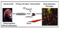
Cellular model systems: Degeneration and cell death occur on a single cell basis asynchronously, which decrease visibility of the alterations on proteome level. To detect and analyse primary alterations leading to cell death, experimental settings have thus to be designed such, that cell death happens synchronously in the majority of all neuronal cells. This can be achieved best ex vivo in primary tissue from normal animals as well as from animals models bearing specific mutations. As most neurodegenerative diseases are regional, we isolate the specific regional tissues affected by disease. With primary tissue, enzymatic cell separation and cell sorting may be necessary to enrich for a cell type to be studied. Forms of stress are then given that synchronously induce apoptosis. We have established in vitro primary ventral-mesencephalic (VM) (Bauer et al., Exp Neurol.,2002) and cortical cell cultures (unpublished), which we can genetically modify by lentiviral gene-transfer. For interference with gene expression, we have established lentivirus mediated RNAi in primary neuronal tissue (Bauer et al., 2005, in revision). We have developed protocols to differentiate rodent progenitor cells and neuronal adult stem cells in vitro into DA neurons. We have developed protocols to challenge cells for synchronized cell death in vitro (Dumont et al., Oncogene1999; Braun et al., submitted). We have extensively analysed and experimentally treated an animal model for Parkinson’s disease, the 6-hydroxy-dopamine lesioned rat (Bauer et al., Hum.Gene Ther 2000; ibid, Neurosci Lett., 2001). We are currently analysing animal models transgenic for disease-associated candidate genes by comparative proteomic analysis (Hauck et al., 2005, Molecular and Cellular Proteomics, submitted).
For rapid experiments and highly standardized experimental conditions, immortalized, yet neurally differentiated PC12 cells offer a convenient and inexpensive option to study physiological and biochemical mechanisms that depend on a uniform cell population and high amounts of material. The functional relevance of candidate proteins potentially involved in the aetiology of disease can be investigated through their recombinant expression or siRNA mediated knock-out in this cell line after neuronal differentiation by Nerve Growth Factor. PC12 cells are then be challenged in a disease-modelling fashion and thereby be driven into synchronized apoptosis.
For genes or proteins whose expression is reduced under pathological conditions, functional complementation assays are performed in which candidate genes are transfected or lentivirally transduced and monitored for their ability to attenuate the defect. PC 12 cells as well as developing brain tissue is primarily being used, as gene-delivery is more efficient and less impairing in these cells. The tissues are then treated such that the pathological condition is present. In complementation to genes or gene variants increasing susceptibility to apoptosis (VCP-, parkin- or LRRK2-variants) candidate neuroprotective genes (neurotrophic factors, certain kinases (PKC, Raf, MAPK) or transcription factors) will be overexpressed and studied for their physiological effect as previously described by us (Grimm et al., 1998; Bauer et al., 2000). On the other hand, identified candidate genes possibly involved in executing or accelerating a pathological condition in brain (for ex.: proapoptotic genes) will be functionally studied by siRNA mediated knock out or targeted by pharmacological means (inhibitors, neuro-protective agents) (Dumont et al., Oncogene,1999).
Proteome analysis: The candidate disease associated genes are first studied through recombinant expression in PC12. If transgenic mice for a given gene are available, primary brain progenitor tissue, using regions affected by the disease will be established. If such mice are not available, primary brain progenitor tissue is virally transduced or transfected with the gene of interest. The genetically modified and eventually challenged tissue material can then be analysed by systematic proteome analysis using 2D-gels (DIGE technology in collaboration with TP1 Meyer) and subsequent mass-spectrometric identification of proteins of interest as published previously (Alge et al., IOVS, 2003; Hauck et al., Glia, 2003, Zischka et al., Proteomics, 2003). Mass-spectrometry of membrane proteins will be facilitated using a novel protocol involving detergent removal by ion-pair extraction (Zischka et al., Proteomics, 2004).
ER and mitochondrial analysis: Endoplasmatic reticulum and mitochondria both shown to be involved in neuronal cell death are being analysed in depth with respect to eventual alterations in protein composition and their role in apoptosis. Using methods for sorting and isolation of subcellular structures established in our group we apply native separation techniques including sucrose density centrifugation and Free Flow Electrophoresis to fractionate the organelles (Zischka et al., Proteomics, 2003; Braun et al., submitted; Isslinger et al., 2005 Proteomics in press) and analyse their functional integrity. Assays for analysing respiratory capacity of mitochondria are established (Zischka et al., in prep.).
Analysis of multiprotein complex composition: Protein complexes are being analysed by affinity purification, native differential fractionation and immunoprecipitation or pull-down, as described within the proposal for SMP Proteomics (TP Ueffing, TP Herberg, TP Meyer) and as previously described by us (Linari et al., 1998, Gires et al., 1999; ibid, 2001).
Valosin containing protein
Proper protein folding, processing and timely turnover of proteins is of key importance for neuronal cell function. Aggregation of misfolded proteins, malfunctions of the ubiquitin-proteasome pathway, due to genetic mutations of proteins involved in this pathway, and ER stress are hallmarks of neurodegenerative disorders. We have investigated the role of Cdc48p, the highly conserved yeast orthologue of valosin-containing protein (VCP/p97). VCP is an effector protein for polyglutamine diseases (Mizuno et al., 2003) and, when mutated, an inductor of “inclusion body myopathy associated with Paget disease of bone and frontotemporal dementia” (IBMPFD), a dominant progressive human disorder (Watts et al., 2004). VCP appears as an important player in the ER-associated protein degradation (ERAD) pathway (Hoppe et al., 2000, Dai and Li, 2001, Ye et al., 2001). It shows increased affinity towards polyubiquitinated proteins and may facilitate their presentation to the proteasome (Rabinovich et al., 2002, Elkabetz et al., 2004). Expression of VCP mutants with impaired functionality result in accumulation and aggregation of membrane-associated polyubiquitinated proteins and ER stress (Kobayashi et al., 2002). From our recent work on mutant CDC48 (Braun et al., 2005 Molecular & Cellular Proteomics, submitted), we propose a direct mechanism of interaction between protein aggregation, ER stress and functional impairment of mitochondria.
Delta-like 1
Delta-like 1 (Dlk1), a member of the Delta/Notch protein family, has been described in rodent developing and mature midbrain dopaminergic (DA) neurons. In the mouse ventral mesencephalon (VM), Dlk1 coincides with tyrosine hydroxylase (TH) expression in Nurr1-positive dopaminergic progenitor cells as early as embryonic day 11.5. We down-regulated Dlk1 protein expression in immature, TH-negative murine VM progenitor cells in vitro using short hairpinRNAs (shRNA) directed against Dlk1 mRNA expressed from a lentivirus vector. shRNA-mediated Dlk1down regulation results in decreased TH mRNA, TH protein and overall numbers of TH-positive neurons in differentiated cultures. The expression of other markers of dopaminergic lineage was also decreased. Dlk1 is therefore likely to contribute to dopaminergic maturation once committed VM progenitor cells begin full differentiation, and may be regarded as a functional marker for full commitment towards mature dopaminergic neurons.
Outlook
An interrelationship of genetic factors and cellular stress has been implicated in several human diseases that have both systemic and neurological implications, including ‘conformational diseases’ such as Huntington's and Parkinson's that are characterized by the accumulation of toxic protein aggregates. (Spiess et al., 2004). Protein aggregation is one of the central pathological hallmarks of neurodegenerative diseases including such prevalent diseases as Alzheimer’s and Parkinson’s but also rare diseases, where no therapeutic strategy has been developed. Identification of the molecular mechanisms of degeneration and cell death associated with Cdc48p/VCP bears implications for a variety of diseases characterized by abnormal protein deposits. Our ongoing study on VCP points at a new, possibly common pathological mechanism of degeneration associated with the ERAD-ubiquitin-proteasome system. Therefore, Cdc48p/VCP-dependent apoptosis may serve as a model system for programmed cell death in human degenerative disorders associated with protein deposits. In addition, its functional interaction with TCP-1 ring complex chaperonins (TRiC, also named CCT for chaperonin-containing TCP1) will be investigated using bioanalytical and cell-biological assays (Braun et al., submitted). Analysing and eventually identifying the systemic consequences of misfolded protein aggregation and its pathophysiological consequences within a cell can contribute to understanding mechanisms of neurodegenerative diseases and offer the opportunity for new therapeutic avenues.In addition to these ongoing analyses we are open to functionally characterize and validate specific genes of interest coming from other partners within HBPP and KG Neuro. We will generate and provide cell culture models that can be used for drug screening and candidate drug validation approaches. In addition, through interactome analysis of disease-associated proteins with their specific interactors we can generate potential candidate genes for association studies. As part of this project we will explore the diagnostic and possible therapeutic value of such targets.
Lit.: 1. Bauer, M., Meyer, M., Brevig, T., Gasser, T., Widmer, H.R., Zimmer J., and Ueffing, M., (2002) Lipid-mediated Glia Cell Line-Derived Neurotrophic Factor Gene Transfer to Cultured Porcine Ventral Mesencephalic Tissue. Experimental Neurology, Exp Neurol. 177:40-49. 2. Hauck, SM, Suppmann, S, and Ueffing, M., (2003) Proteomic profiling of primary retinal Müller glia cells reveals a shift in expression patterns upon adaption to in vitro conditions. Glia,44, 251-63