


Introduction
With the complete human genome sequence available (1) we now need functional genomics methods to discover protein function genome-wide. RNA interference (RNAi) is the method of choice to study loss of function phenotypes in human cells because it allows to specifically suppress the expression of virtually any desired protein-coding gene. In human cells, the cost of genome-wide libraries of chemically synthesized 21mers (siRNAs) which represent the best validated RNAi reagent for mammals (2) has so far limited their use to targeted screens of several hundred genes (3, 4). For the alternative technology of short hairpin RNAs larger libraries are available and have been used in simple assays (5-7), but shRNAs require six or more reagents per gene. Finally, cocktails of interfering RNAs generated by enzymatic digestion of long double stranded molecules (esiRNAs) have been produced as large libraries and again used in simple assays (8).
Fluorescence microscopy provides a uniquely detailed phenotypic readout of cultured cells after RNAi suppression of a gene (9). A key technology to achieve this in a cost efficient manner are RNAi microarrays which can locally transfect hundreds of siRNAs printed on glass slides into overlaid cells and are thus referred to shortly as transfected cell arrays (10-12). Combined with automated microscopy (13-14) cell arrays allow the detailed analysis of cellular phenotypes after RNAi knock-down in high throughput. The aim of this subproject is therefore to integrate recent advances in mammalian cell transfection and microscopy automation into a platform for genome-scale RNAi screens in human cells. By using large RNAi libraries cost efficiently for high content and high throughput screening, genome-wide analysis of gene function has the potential to become a routine technology for human cell systems. By applying this technology to assays relevant for cancer and neuronal diseases, we aim to identify gene networks required for biological processes central to human diseases.
Project Status
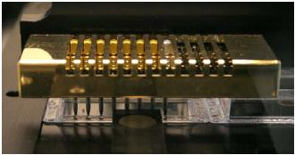
Fig 1: Printing of siRNA microarrays are into live cell imaging chambers.
This project has started in May 2005 and is therefore still in the initial technology development phase. The first step to set up a platform for high throughput genome-scale RNA interference screens in cultured human cells was the optimization and production of transfected cell arrays. This technology has been developed successfully for siRNAs with high transfection efficiencies for HeLa cells and currently densities of 384 siRNAs/live cell imaging chamber are reached by robotic deposition of siRNA transfection reagent cocktails (Fig. 1). Optimization of this method for reverse transfection for esiRNAs is ongoing.
The next step in the workflow is the analysis of cellular phenotypes by automated fluorescence microscopy. To this end, we are currently setting up a fully automated confocal screening microscope to increase throughput of assays requiring confocal resolution. In parallel we are developing appropriate high throughput phenotypic assays based on reporter cell lines expressing fluorescent proteins for mitosis (relevant for cancer), microtubule stability (relevant for neuronal diseases) and secretion (relevant for Alzheimer and related diseases). Assay development for mitosis and secretion are well advanced while microtubule stability is still ongoing. The first screening experiments for the secretion assay have been started.
Outlook
After completion of technology development, we will perform genome-scale RNAi screens for genes required for cell cycle progression, protein secretion and microtubule stability. These screens will deliver phenotypic fingerprints from each assay in an unbiased fashion for most genes in the human genome. The genes identified in this fashion will be subjected to further functional analysis both in this project, by tagging them with fluorescent and affinity tags and stably expressing them in human cell lines, as well as in other projects of this SMP in the more physiological systems of embryonic and adult mice.
Lit.: 1. International Human Genome Sequencing Consortium. Finishing the euchromatic sequence of the human genome. Nature. 2004 431(7011):931-45. 2. Elbashir SM et al. Duplexes of 21-nucleotide RNAs mediate RNA interference in cultured mammalian cells. Nature 2001 411(6836):494-8. 3. Aza-Blanc P et al. Identification of modulators of TRAIL-induced apoptosis via RNAi-based phenotypic screening. Mol Cell. 2003 12(3):627-37. 4. Pelkmans L et al. Genome-wide analysis of human kinases in clathrin- and caveolae/raft-mediated endocytosis. Nature. 2005 Jul 7;436(7047):78-86. 5. Zheng L et al. An approach to genomewide screens of expressed small interfering RNAs in mammalian cells. Proc Natl Acad Sci USA. 2004 101(1):135-40. 6. Berns K. et al. A large-scale RNAi screen in human cells identifies new components of the p53 pathway. Nature. 2004 428(6981):431-7. 7. Paddison PJ. et al. A resource for large-scale RNA-interference-based screens in mammals. Nature. 2004 428(6981):427-31. 8. Kittler R et al. An endoribonuclease-prepared siRNA screen in human cells identifies genes essential for cell division. Nature. 2004 432(7020):1036-40. 9. Goshima G. and Vale RD. The roles of microtubule-based motor proteins in mitosis: comprehensive RNAi analysis in the Drosophila S2 cell line. J Cell Biol. 2003 162(6):1003-16. 10. Erfle H et al. siRNA cell arrays for high-content screening microscopy. Biotechniques. 2004 37(3):454-8, 460, 462. 11. Silva JM et al. RNA interference microarrays: High-throughput loss-offunction genetics in mammalian cells. Proc Natl Acad Sci USA. 2004 Apr 27;101(17):6548-52. 12. Ziauddin J and Sabatini DM. Microarrays of cells expressing defined cDNAs. Nature. 2001 411(6833):107-10. 13. Liebel U et al. A microscope-based screening platform for large-scale functional protein analysis in intact cells. FEBS Lett. 2003 554(3):394-8. 14. Starkuviene V et al. High-content screening microscopy identifies novel proteins with a putative role in secretory membrane traffic. Genome Res. 2004 14(10A):1948-56.