


Introduction
Tumor-specific genetic aberrations are not only important for the understanding of the molecular mechanisms underlying the development of cancer, but can also provide important diagnostic and prognostic markers, which are useful for optimal therapy decisions. DNA/cDNA based array technologies are highly efficient tools for a genome-wide screening of such aberrations. However, the number of cases from individual tumor entities, which can be analyzed within reasonable time and cost limits usually remain <100. Since in most instances, this is not sufficient to determine genotype/phenotype correlations by robust statistical testing, such analyses have to be performed on larger tumor collections. For this, the approach of tissue microarrays (TMAs) can be efficiently used.
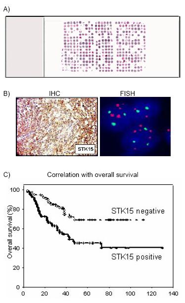
Fig 1: Combined DNA- and tissue-chip analysis to identify novel diagnostic and predictive molecular markers. (A) Example of a TMA containing ~450 biopsy cores of head and neck squamous cell carcinoma (HNSCC). (B) Example for an IHC (left) and a FISH experiment (right) on TMA tissue cores derived from medulloblastoma. The IHC experiment shows tumor tissue positive for aurora kinase A (STK15) involved in the control of genomic stability. The FISH experiment shows additional copies of the STK15 gene. (C) Correlation of increased expression of STK15 with unfavourable overall survival (4).
We have generated a large number of TMAs, while special emphasis was drawn on the quality of the tumor material, which was characterized by expert pathologists in all cases. Furthermore, for most of the TMAs, survival parameters in addition to a detailed patho-morphological characterization is available. Analysis of these arrays therefore allow to rapidly correlate cytogenetic changes or aberrant protein expression with important clinical parameters.
Results/Project Status
The role of Cyclins in Squamous Cell Carcinoma of the Head and Neck (HNSCC): HNSCC represent the 6th most frequent tumor type world wide, but despite of modern therapeutic strategies like adjuvant and neoadjuvant radio- and chemotherapy, the overall 5-year survival rate does not exceed 55%. Furthermore, HNSSCs are a quite heterogeneous group presenting as tumors of different anatomical regions such as the oral cavity, the oro-/hypopharynx and the larynx, which show a different clinical behaviour. In collaboration with Ch. Hofele and F.X. Bosch (Heidelberg), a major interest in this project was to analyse the role of the CyclinD1 gene during HNSCC development (1,2). We detected CyclinD1 amplifications in 35% on our TMA containing ~500 different tumors. The pharyngeal site was preferentially affected, which was in concordance with further protein measurement studies. These data further supported, that HNSCC of different localization follow different pathological pathways. An interesting cytogenetic aberration in HNSCCs are copy number gains of chromosomal subregion 3q25-q29, since this has been associated with shortened disease-specific survival. We tested several putative oncogenes from this region (SNO, PIK3CA, TP73L, CyclinL1) (3). Most interesting results were obtained for CyclinL1 (Fig.2), since copy number gains of this gene were found significantly associated with the presence of lymph node metastases. Furthermore, Kaplan�Meier analysis revealed, that high-level amplifications of CCNL1 correlated with shorter overall survival. Thus, beside CyclinD1 also CCNL1 plays a critical role in the progression of HNSCC and may serve the clinicians as an important indicator for occult metastases.
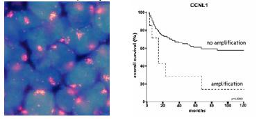
Fig 2: Analysis of Cyclin L1 in HNSCC tumors. Left: FISH analysis showing nuclei (blue) of tumor cells harbouring a high-level amplification of CyclinL1 (red); Right: Kaplan-Meier plot indicating an unfavourable clinical course in patients with CyclinL1 amplification (3).
Analysis of anti-apoptotic proteins in Breast- and Colorectal Carcinoma: In colaboration with M. Zörnig (Frankfurt) we test newly defined anti-apoptotic proteins, identified by functional yeast survival screens, for their potential diagnostic relevance in different tumor systems. Among others, expression of the high-mobility group box-1 protein (HMGB1) as well as the c-IAP protein, which becomes upregulated by HMGB-1, were studied by means of TMAs with breast- and colon carcinomas (5,6). In both tumor types, the expression level was found significantly increased within the tumor cells. For quantitative measurement of the protein expression, we used an approach, which we developed to allow for automated image aquisition and evaluation using fluorochrome-labeled antibodies. This is shown for c-IAP expression in colon carcinomas in Fig. 3.
Adenoid Cystic Carcinoma: Novel therapeutic interference points? Adenoid cystic carcinoma (ACC) is the second most common malignant tumor of the salivary glands and characterized by a prolonged but inevitably unfavorable clinical course. Recent studies suggested the transmembrane tyrosine kinase KIT to be involved in ACC pathogenesis. To investigate KIT expression in histologically defined subgroups of ACC and to clarify whether KIT gene copy number gain contributes to KIT overexpression, TMA sections including 55 ACC tumors were analyzed (7). Gene amplifications of KIT were detected in 6%, while overexpression of the protein was detected in 89% of the cases. Kit was subtype-specifically expressed in cribiform and tubular ACCs, but never in solid subtypes. These results implicate, that specific KIT tyrosine kinase inhibitors such as imatinib, might be used in future therapeutic approaches against ACC. We also used the ACC-TMA to identify the critical target genes affected by frequent chromosome 22 copy number gains found in ACC (8).
Expression of BCL3 in Malignant Lymphoma and Hodgkin´s Lymphoma: In collaboration with H. Stein (Berlin) we constructed TMAs from malignant lymphoma including 48 T cell derived lymphomas, 72 cases of classical (cHL) and lymphocyte predominant Hodgkin´s lymphoma (LPHL) as well as 164 B-cell derived lymphomas. Using these TMAs we recently analyzed the expression of the antiapoptotic oncoprotein BCL-3 in the different lymphomas (8). Applying IHC analysis, we observed a strong Bcl-3 expression in cHL and in peripheral T-cell lymphomas including anaplastic large cell lymphomas (ALCL). Furthermore, we detected in 3/6 cHL cell lines and 25% of primary ALCL a copy number increase of the Bcl-3 gene locus. These results suggest that elevated Bcl-3 expression has an important function in cHL and peripheral T-cell lymphomas, in particular ALCL.
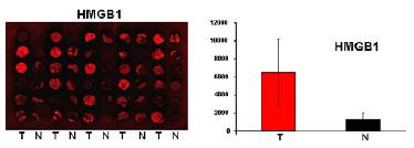
Fig 3: Analysis of TMAs using fluorochrome-labelled antibodies. (A) HMGB-1-immunostaining of a TMA with biopsies from primary colon carcinoma (T) and normal colon tissue (N). The digital image was obtained by means of a scanning device, which is currently developped in collaboration with Carl Zeiss Jena GmbH. (B) Quantitative measurement of the average fluorescence intensity of HMGB-1 within the different biopsy cores (6).
Beside the analyses described above, we generated and analyzed microarrays from CNS tumors (in collaboration with the NGFN2-Glioma Network coordinated by O.D.W. Wiestler, Heidelberg) (9) as well as myocardiac tissue samples (in collaboration with the NGFN2 network for cardio-vascular diseases; G. Hasenfus and R. Knoell, Göttingen). We will continue to generate high-quality TMAs, in particular from colorectal carcinomas. Furthermore, we want to concentrate on the analysis of cell signalling proteins as well as antiapoptotic proteins. Finally, we focus on the further development of a suitable automated image acquisition and evaluation system in order to make TMA analyses still more efficient for the search of clinically important markers.
Lit.: Freier K et al. Tissue microarray analysis reveals site-specific prevalence of onogene amplifications in head and neck squamous cell carcinoma. Cancer Res. 2003; Mar 15;63(6):1179-82. 2. Freier K et al. Distinct site-specific oncoprotein overexpression in head and neck squamous cell carcinoma: A tissue microarray analysis. Anticancer Res. 2003; Sep-Oct 23(5A):3971-7. 3. Sticht C et al. Amplification of CyclinL1 is associated with lymph node metastases in head and neck squamous cell carcinoma (HNSCC). Br J Cancer. 2005 Feb 28;92(4):770-4. 4. Neben K et al. Microarray-based screening for molecular markers in medulloblastoma revealed STK15 as independent predictor for survival. Cancer Res. 2003; May 1; 64(9), 3103-11 5. Brezniceanu ML, et al. HMGB1 inhibits cell death in yeast and mammalian cells and is abundantly expressed in human breast carcinoma. FASEB J, 2003,17(10):1295-7. 6. Völp K et al. Expression of high mobility group box 1 (HMGB1) is associated with an elevated level of the anti-apoptotic c-IAP2 protein in human colon carcinomas. Gut, 2005, in press.7. Freier K et al. Differential KIT protein expression in histological subtypes of adenoid cystic carcinoma (ACC) of the salivary gland. Oral Oncology 2005, Jul 26; Epub ahead of print. 8. Freier K et al. Novel copy number gains on chromosome 22q13 in adenoid cystic carcinoma (ACC) of the salivary gland revealed by comparative genomic hybridization (CGH) and tissue microarray (TMA) analysis Cytogenetics and Cell Genetics, 2005 May;159(1):89-95. 9. Mathas S et al. Elevated Bcl-3 expression and p50 complex formation in anaplastic large cell, classical Hodgkin- and peripheral T cell lymphomas, Blood, 2005, in press.